How Does Breast Cancer Look Like On A Mammogram / Mammogram Images Normal Abnormal And Breast Cancer : The outer edges of these cells look fuzzy or spiky (called spiculated).
Dapatkan link
Facebook
X
Pinterest
Email
Aplikasi Lainnya
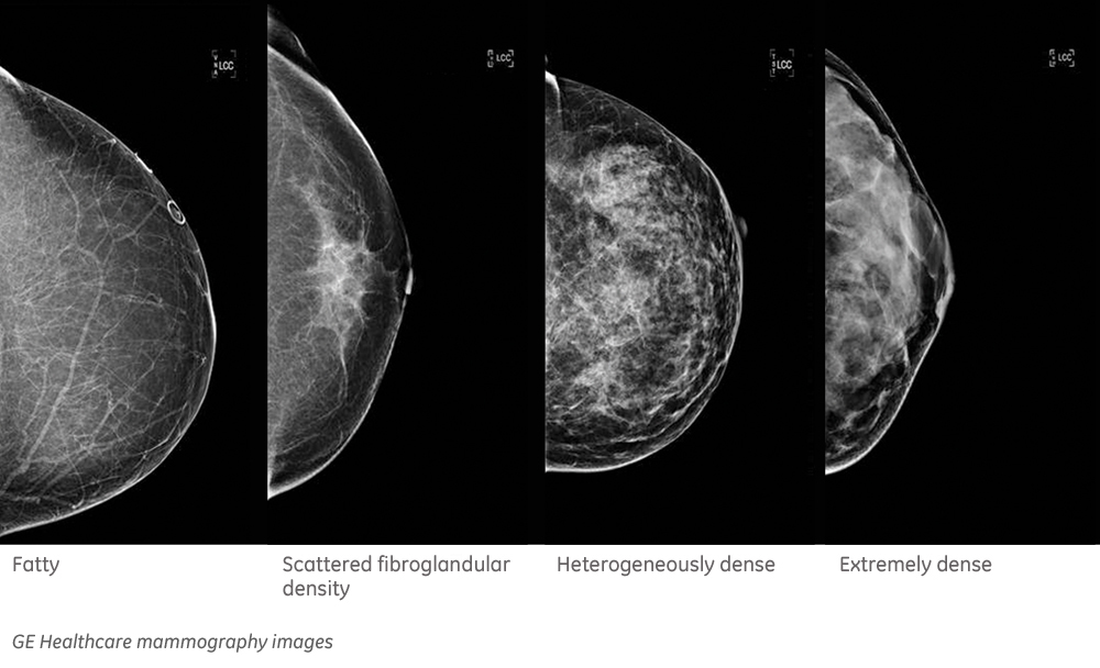
How Does Breast Cancer Look Like On A Mammogram / Mammogram Images Normal Abnormal And Breast Cancer : The outer edges of these cells look fuzzy or spiky (called spiculated).. Healthy mammograms can still vary in appearance. That makes it easy to detect abnormalities, which generally show up as white. If found in an area of rapidly dividing cells or grouped together in a certain way, they may be a sign of dcis or breast. A lump or tumor will show up as a focused white area on a mammogram. Dense breast tissue appears solid.
Breast cancer and some noncancerous (benign) breast conditions can appear white on a mammogram. A breast mri captures multiple images of your breast. A 3d mammogram is used to look for breast cancer in people who have no signs or symptoms. What does cancer look like on a mammogram? A rash isn't the only visual symptom of inflammatory breast cancer.
Breast Masses Cancerous Tumor Or Benign Lump from www.verywellhealth.com Even if you have a lump in only one breast, pictures will be taken of both breasts. This appears most commonly as streaking, known as linear enhancement. A woman's breast tissue also changes over time, and it is not uncommon for benign lumps, cysts or calcifications to form with age. Breast cancer can appear as a spiculated mass, cluster of tiny calcifications, smoothly marginated mass, area of subtle distortion or be invisible on. What does an abnormal mammogram look like? Cancer cells can remain within the milk ducts and this is considered as noninvasive cancer or ductal carcinoma in situ. Magnetic resonance imaging (mri) of the breast — or breast mri — is a test used to detect breast cancer and other abnormalities in the breast. The appearance of normal breast tissue on a mammogram varies from person to person, and no two mammograms look the same.
Dense breast tissue appears solid.
Any area that does not look like normal tissue is a possible cause for concern. Magnetic resonance imaging (mri) of the breast — or breast mri — is a test used to detect breast cancer and other abnormalities in the breast. Breast cancer and some noncancerous (benign) breast conditions can appear white on a mammogram. Microcalcifications, which look like white specks on a mammogram. What does an abnormal mammogram look like? Breast mri images are combined, using a computer, to create detailed pictures. Mammograms may show suspicious areas of the breast, white spots, and microcalcifications. In addition to mammograms, ultrasound and mri may also be used to take a closer look at changes in the breast. Tumors are likely to be smaller when doctors detect them early, which can. This type of cancer also changes the appearance of your breasts. A lump or tumor will show up as a focused white area on a mammogram. This appears most commonly as streaking, known as linear enhancement. There are few risks associated with mammography.
Abnormalities such as cancerous tumors usually appear brighter because they are denser. If found in an area of rapidly dividing cells or grouped together in a certain way, they may be a sign of dcis or breast. They are often caused by aging, an old injury, or inflammation and are usually benign. A breast mri usually is performed after you have a. A breast mri captures multiple images of your breast.
Breast Cancer Ge News from www.ge.com Healthy mammograms can still vary in appearance. The dye collection in the breast can also look clumpy or appear in a section of the breast, depending on the involvement of dcis. It can also be used to investigate the cause of breast problems, such as a breast mass, pain and nipple. Cancers may be seen as masses (like a ball, but usually with an irregular shape), areas of asymmetry that resemble normal tissue, calcifications (white specks), and/or areas of architectural distortion (imagine the puckering caused by pulling a thread in a piece of fabric). There are few risks associated with mammography. If you've had a mammogram before, the radiologist should compare your old mammogram to the new one to look for changes. After a mammogram that didn't show anything, and a sonogram that found the lump, i was diagnosed with stage 2 breast cancer. The doctor reading your mammogram will be looking for different types of breast changes, such as small white spots called calcifications, larger abnormal areas called masses, and other suspicious areas that could be signs of cancer.
Abnormalities such as cancerous tumors usually appear brighter because they are denser.
Microcalcifications, which look like white specks on a mammogram. They will look carefully at the mammogram to interpret the results. Tumor size is an important factor in breast cancer staging, and it can affect a person's treatment options and outlook. Breast mri images are combined, using a computer, to create detailed pictures. Breast cancer can appear as a spiculated mass, cluster of tiny calcifications, smoothly marginated mass, area of subtle distortion or be invisible on. Magnetic resonance imaging (mri) of the breast — or breast mri — is a test used to detect breast cancer and other abnormalities in the breast. Calcifications are calcium deposits within the breast tissue and they look like small white spots. A breast cancer tumor on a mammogram is often irregular with edges that don't look smooth. A 3d mammogram is used to look for breast cancer in people who have no signs or symptoms. A rash isn't the only visual symptom of inflammatory breast cancer. A mammogram can show breast changes such as calcifications, masses, or other symptoms that might be cancer. This appears most commonly as streaking, known as linear enhancement. What does cancer look like on a mammogram?
This appears most commonly as streaking, known as linear enhancement. As with all abnormalities seen on breast imaging, the diagnosis of dcis requires a sample of tissue or biopsy. What does breast cancer look like on a mammogram? Calcifications are calcium deposits within the breast tissue and they look like small white spots. Breast cancer can appear as a spiculated mass, cluster of tiny calcifications, smoothly marginated mass, area of subtle distortion or be invisible on.
Mammogram Images Normal And Abnormal from www.verywellhealth.com Any area that does not look like normal tissue is a possible cause for concern. The appearance of normal breast tissue on a mammogram varies from person to person, and no two mammograms look the same. A woman's breast tissue also changes over time, and it is not uncommon for benign lumps, cysts or calcifications to form with age. What does cancer look like on a mammogram? Tumors may be benign or cancerous. Abnormalities such as cancerous tumors usually appear brighter because they are denser. Cancers may be seen as masses (like a ball, but usually with an irregular shape), areas of asymmetry that resemble normal tissue, calcifications (white specks), and/or areas of architectural distortion (imagine the puckering caused by pulling a thread in a piece of fabric). Calcifications are calcium deposits within the breast tissue and they look like small white spots.
Cancers may be seen as masses (like a ball, but usually with an irregular shape), areas of asymmetry that resemble normal tissue, calcifications (white specks), and/or areas of architectural distortion (imagine the puckering caused by pulling a thread in a piece of fabric).
Any area that does not look like normal tissue is a possible cause for concern. Invasive breast cancer can appear as a white patch or mass on a mammogram. You may notice dimpling or pitting, and the skin on your breast. A rash isn't the only visual symptom of inflammatory breast cancer. The milk ducts carry your breast milk from lobules, where milk is produced, to your nipple. Magnetic resonance imaging (mri) of the breast — or breast mri — is a test used to detect breast cancer and other abnormalities in the breast. A lump or tumor will show up as a focused white area on a mammogram. 1 the gray areas correspond to normal fatty tissue, while the white areas are normal breast tissue with ducts and lobes. A spiculated breast mass, which has spikes extending out from the main mass, is often highly suggestive of cancer. Macrocalcifications, which look like small white dots on a mammogram. It's so important to listen to the messages our bodies are telling. In addition to mammograms, ultrasound and mri may also be used to take a closer look at changes in the breast. Breast cancer can appear as a spiculated mass, cluster of tiny calcifications, smoothly marginated mass, area of subtle distortion or be invisible on.
Syarat membuat surat perjanjian jual beli rumah. Akta pendirian koperasi simpan pinjam; Warisan, hibah, hibah wasiat dan wakaf. S e b e l u m d o w l o a d b a c a d u l u : Didalam syara sendiri menyebutkan hibah mempunyai arti akad pokok persoalannya pemberian Surat Persetujuan Kredit from imgv2-1-f.scribdassets.com Semua kekayaan lembaga harus dipergunakan untuk mencapai dan. Surat wasiat harus kamu buat sesuai dengan baik agar informasinya jelas. Warisan, hibah, hibah wasiat dan wakaf. Akta pendirian koperasi simpan pinjam; 06.09.2021 · tak hanya tanah, hibah juga dapat berupa bangunan, rumah, atau barang berharga lainnya. 09.12.2015 · dibawah ini terdapat draft dari beberapa akta notaris (baik partij akte maupun relaas akta), tentunya sebagai sebuah draft, ini bukanlah menjadi akta yang baku namun hanya digunakan sebagai pembelajaran ...
Apart green palace tower mawar lantai hokki 8 sedang unfurnished istimewah shm cash di luar biaya ajb 2 pajak jual/beli balik nama shm dan notaris ya … Hal ini disampaikan luhut dalam konferensi pers melalui kanal youtube sekretariat … 500 2 lantai kamar tidur : Sementara nomor hak adalah nomor sertifikat tanah yang dapat dilihat di halaman dua pada sertifikat hak milik (shm) atau sertifikat hak guna bangunan (shgb), tepatnya di bawah gambar garuda. Sumur/jet pump shm harga : DEALER DAIHATSU SURABAYA | DAIHATSU SURABAYA | SHOWROOM from dealerdaihatsusurabaya.com Booming juga turut mempengaruhi kenaikan harga satu jenis properti yang sedang banyak diincar. Carport garden garasi refrigerator ac pam swimming pool stove fire extenguisher telephone. + free bphtb + free canopy + free indihome 6 bulan + free balik nama + free notaris + free pnbp ...
How To Make Internal Storage To Sd Card / How To Use Sd Card As Internal Storage On Android Adoptable Storage On Android : I have added a pen drive to act as external storage 2. . .storage to sd card so that you can easily free up the internal storage of your device and make without any further ado, let us have a look at how to move apps from the internal storage of a the good news is that it is very straightforward to move apps from internal storage to sd card on android. Make sure all the data on the sd now that you have learnt how to format and reformat your microsd card to use as internal. But then how do you format your sd card for use as extra internal storage? Let you install huge apps directly in the sd cards. How to set micro sd. So on that condition we can do one thing and that is swap make sure you changes right values, if not then you might brick your phone or sd card. Sd card is widely used in portable devices, like digital. The differences between porta...
/breast-cancer-tumors-what-are-they-430277-v12-d91aad27f20b4f06aae6afc5a55868da.png)
:max_bytes(150000):strip_icc()/calcifications-56a0b6835f9b58eba4b30bc3.jpeg)


Komentar
Posting Komentar